
Gastrointestinal Anatomy and Physiology of Ruminants
LPM-00500 View this publication in PDF form to print or download.
by Joanne McCrea
Ruminants are a group of animals that includes cattle, goats, sheep and reindeer. These animals have a unique digestive system with one stomach divided into four parts: the rumen, the reticulum, the omasum and the abomasum. The largest stomach is the rumen, which acts like a fermentation vat where food is broken down by microbes (bacteria, protozoa and fungi). The microbial fermentation process allows ruminants to transform tough, fibrous plants into the energy and nutrients needed to produce important food products for humans, such as milk and meat.
Through fermentation, microbes inside the rumen break down food into volatile fatty acids (VFAs) and other nutrients. These volatile fatty acids are absorbed through the rumen and intestinal wall into the bloodstream and help the animal produce sugars and fats. The main types of volatile fatty acids produced are lactic, acetic, propionic and butyric acids.
The microbes in the rumen also create B vitamins, vitamin K and amino acids. Amino acids are the building blocks of proteins that contain a large amount of nitrogen. The microbes can use nonprotein nitrogen sources to build proteins that can be digested and used by the animal. Good management practices that support a healthy rumen environment are crucial forkeeping ruminants healthy and productive.
Ruminants also engage in a behavior called rumination, or chewing cud. This involves regurgitating partially digested food from the rumen, chewing it and swallowing it again. This process helps further break down the long fibers in their food.
When ruminating, the animals produce more saliva, which helps maintain a healthy environment in the rumen and supports microbial fermentation. Typically, a ruminant will spend six to eight hours a day chewing cud, usually while calm and lying in a sternal position (a position where an animal lies on its sternum, or breastbone, with its back facing upward). However, stress or other factors can reduce the time they spend chewing cud or grazing.
Knowledge of ruminant gastrointestinal anatomy and physiology can help livestock owners understand risk factors, causes, prevention and treatment of common gastrointestinal disorders.

Head and Neck region
The head and neck contain several important structures, including the lips, mouth, tongue, dental pad, teeth, salivary glands, throat (pharynx) and esophagus.
The lips, tongue, lower incisors, and dental pad of the upper jaw collect food. The premolar and molar teeth of the upper and lower jaws chew and grind food into smaller pieces so it can be swallowed and digested. The tongue moves food around in the mouth and aids in swallowing.
In ruminants, the salivary glands produce a lot of saliva that moistens the food and adds important substances like sodium, potassium and bicarbonate to the rumen. Bicarbonate has a basic pH. It helps neutralize acids produced by microbial fermentation, thereby maintaining a normal rumen pH and a healthy rumen environment.
Urea is a byproduct formed when protein is broken down during metabolism. It is produced in the liver, transported to the kidneys and excreted in urine. It is also found in the saliva. While animals don’t use the nitrogen found in urea directly for making proteins, it can be used by microbes in the rumen to create microbial protein. This protein is later digested in a part of the stomach called the abomasum and absorbed in the small intestine. This is a form of urea recycling that allows ruminants to conserve the nitrogen from protein intake.
The pharynx is the region located at the back of the mouth where the mouth connects to the esophagus and the nasal passages connect to the larynx (voice box) and trachea. Coordinated muscle movements of this region enable swallowing. The larynx or voice box is also in this region and includes the epiglottis, a door-like flap that covers the airway during swallowing and regurgitation to prevent food and fluids from entering the trachea.
The esophagus is a tube that connects the mouth to the forestomach. Swallowed food and water travel down the esophagus into the forestomach. Food material in the rumen will be regurgitated from the rumen into the esophagus. Gas produced during digestion will leave the rumen through the esophagus in a process called eructation (or belching). This is crucial for preventing gas buildup in the rumen, which can be uncomfortable and life-threatening for the animal.
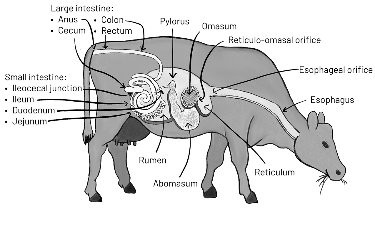
Ruminant Stomach
The stomach of ruminant animals has four compartments: the reticulum, rumen, omasum and abomasum. The first two parts, the reticulum and rumen, are often called the reticulo-rumen because there is only partial separation between the two compartments. Inside the forestomach, there are also important openings that allow for the movement of food between regions. The esophageal orifice is where the food from the esophagus enters the upper part of the reticulum. Then, there’s the reticulo-omasal orifice, which is the passage that lets small bits of food and liquid move from the reticulo-rumen into the omasum. Finally, the pylorus connects the end of the abomasum to the start of the small intestine. The pylorus is a narrow and thick-walled structure through which food passes. This narrow passageway can sometimes become blocked.
Reticulum and rumen (reticulo-rumen)
The reticulum is the first part of a ruminant’s forestomach. It is connected to the rumen, the second compartment, but not completely separated, allowing material being digested to flow between these compartments. Because of this, the two are often referred to together as the reticulo-rumen.
The main job of the reticulum is to sort and transport food to the rumen, esophagus or omasum (the third compartment). When food comes in from the esophagus, lightweight or large pieces go into the rumen, while heavier items like nails or wire sink to the bottom of the reticulum. During regurgitation, the rumen contents come back through the reticulum and the esophagus into the animal’s mouth. Smaller food particles that have already been broken down pass through the reticulo-omasal orifice for further digestion in the omasum and abomasum.
The rumen is the largest part of the forestomach and is where microbial fermentation occurs. It is divided into different sacs by muscular pillars. These pillars provide structural support for the rumen and help with rumen motility — the rhythmic, coordinated muscular contractions of the rumen and reticulum. These contractions enable regurgitation, belching and the mixing of rumen contents. The internal lining of the rumen has tiny finger-like projections called papillae that increase the surface area for absorbing nutrients. The rumen lining is a selective barrier that allows absorption of some nutrients, like volatile fatty acids, into the animal’s bloodstream.
Rumen motility can be divided into primary and secondary rumen contractions. Primary rumen contractions occur about once to three times a minute and help mix the food. During this mixing process, smaller, heavier particles sink to the bottom while lighter, fibrous pieces float to the top. Gases produced from fermentation also affect the density of the food that is not fully digested, allowing it to float. The smaller particles then move into the omasum, while larger fibrous pieces are regurgitated for more chewing. Regurgitation helps break down the food further, increasing its surface area, which allows for more efficient fermentation. Primary rumen contractions also increase the amount of volatile fatty acids absorbed into the bloodstream by causing more contact with the rumen papillae.
Secondary contractions help release gas via belching. These contractions push a pocket of gas from the top of the rumen into the reticulum and up the esophagus.
Milk bypass and rumen development in young animals
In young ruminants, milk normally bypasses the reticulo-rumen and goes directly into the omasum. This happens through a special groove called the reticulo-omasal groove, or esophageal groove, a channel of two muscular folds within the reticulum that, when closed, connects the esophagus to the omasum. When a healthy young animal suckles, nerve signals cause the groove to close, forming a tube that allows milk to flow past the reticulo-rumen and into the omasum. If this groove doesn’t close properly, serious infections and digestive problems can occur.
Young animals’ rumen and rumen papillae develop over the first few months of life. Eating solid food and establishing normal rumen microbes are important for this development.
Omasum
The omasum is smaller than the other stomach compartments, but its interior has many folded sheets that increase the surface area for absorbing water from the food. Its main job is to reabsorb fluids. Due to its appearance of stacked sheets, the omasum has earned it the nickname “book” or “leaf stomach.”
Abomasum
The abomasum is often called the true stomach because, like the stomach of a monogastric animal (such as a human or pig), it contains glands that produce hydrochloric acid for digestion. The abomasum also secretes an enzyme called lysozyme, which helps break down bacteria that enter the abomasum, allowing the nutrients from those bacteria to be absorbed in the small intestine.
Intestines
Small intestine
After the abomasum, food moves into the small intestine, where most nutrients are absorbed through the intestinal wall and into the bloodstream. The small intestine has three sections: the duodenum, jejunum and ileum. Food from the abomasum exits through the pylorus and enters the duodenum, where enzymes from the pancreas and bile from the liver are mixed with the food. Pancreatic enzymes and bile are needed for the chemical reactions involved in digesting and absorbing nutrients. The jejunum is the longest section of the small intestine and connects to the shorter ileum, which then connects to the cecum of the large intestine at a point called the ileocecal junction.
Large intestine
The large intestine, or large colon, absorbs a small amount of nutrients, but its primary role is to absorb water. The large intestine is made up of the cecum, colon and rectum. The cecum is a pouch between the small intestine and the colon and provides some microbial fermentation and water absorption. Ruminants have a three-part colon: spiral, transverse and descending colon. This is where feces are formed. The rectum is the final part of the large intestine and connects to the anus.
Liver and pancreas
The liver is a vital organ that detoxifies the blood, produces proteins and creates bile — a substance that helps digest fats. Bile is stored in the gallbladder for later use and is released into the duodenum through the bile duct when needed.
The hepatic portal vein is a large vein that carries blood from the stomach, small intestine, large intestine, spleen and pancreas to the liver. The liver processes these nutrients into forms that the animal can use. If there’s damage to the rumen or intestinal lining, bacteria may enter the bloodstream, hepatic portal vein and liver. If there are too many bacteria for the liver to detoxify, infection may occur, and bacteria may spread through the bloodstream.
The pancreas is a gland located next to the duodenum that releases enzymes that aid in the digestion and absorption of nutrients. These enzymes travel through a duct that connects to the bile duct, which enters the duodenum.
Joanne McCrea, Assistant Professor of Large Animal Medicine
New March 2026